. 2020 Dec 30;9:e60432. doi: 10.7554/eLife.60432

Figure 9. Rock and Roll is affected in the core planar cell polarity (cPCP) mutant, trilobite.

(A) Frequencies of Roll and No Roll of nascent hair cell (HC) pairs in control (vangl2m209/+; emx2gfp/+) and mutant (vangl2m209/m209; emx2gfp/+) larvae. Significance was assessed by using chi-squared test with a 2 × 2 contingency table (X2[(df = 1)]=6.1774, p<0.013, source data 1). (B–D) Duration of Rock (B), Roll (C), and Rock and Roll (D) of sibling HCs that underwent Rock and Roll in control (n = 28 from seven larvae) and vangl2m209/m209; emx2gfp/+ (n = 19 from seven larvae) larvae. Significance was assessed by using Student's t-test (*p<0.05, **p<0.001, source data 2). The error bars represent SEM. (E) Time-lapse images of a HC pair in trilobite (vangl2m209/m209); emx2gfp/+ mutant, which undergoes a prolong Roll phase from 78 to 246 min with several rolls (first roll: 78–96 min, second roll: 102–120 min, third partial roll: 138–162 min) and resulted in the emx2:Gfp-positive HC located to the posterior. The following figure supplement, source data, and video are available for Figure 9—figure supplement 1. Formation of apical protrusion in nascent HCs of trilobite.

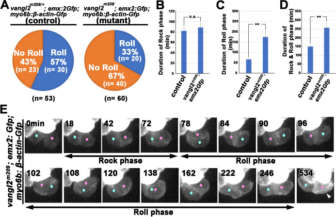
Figure 9—source data 1. Frequencies of Roll and No Roll movements in vangl2 mutants.
Figure 9—source data 2. Comparison of duration of Rock, Roll, and R and R between controls and vangl2 mutants.

Figure 9.

Figure 9—figure supplement 1. Formation of apical protrusion in nascent hair cells (HCs) of trilobite.

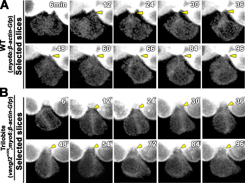
Figure 9—figure supplement 1.

Time-lapse images of a nascent HC pair during Rock phase in (A) wild-type (WT) (myo6:β-actin-Gfp) and (B) trilobite larvae. The apical protrusion (yellow arrowhead) was evident by 12 min after the HC precursor divided in both WT and mutant.
Figure 9—video 1. Time-lapse video of a vangl2m209; emx2: Gfp; myo6b: β-actin-Gfp hair cell (HC) pair during Rock and Roll in Figure 9E.
Download video file (1.3MB, mp4)
The HC precursor (yellow dot) divides to form two sibling HCs (pink and blue dots), which underwent the Rock movement (0–72 min) and then rolled to exchange positions with each other (78–96 min). After the first exchange, the sibling HCs underwent another exchange (102–246 min). By the end of the Roll phase, the emx2 reporter-positive HC (pink dot) was mispositioned at the posterior position, as evident by its brighter GFP signal, compared to the emx2 reporter-negative HC (blue dot) located in the anterior position (534 min).