. 2020 Dec 14;9:e62614. doi: 10.7554/eLife.62614

Figure 1. DolP is a conserved BON-domain protein with a distinct role in OM homeostasis.

(A) In E. coli, dolP is located downstream of diaA and encodes a lipoprotein with a signal sequence (orange) and two BON domains (red). The signal sequence is cleaved by LspA, the cysteine at position 19 acylated by Lgt and Lnt and finally the protein is targeted to the OM by the Lol system (Figure 1—figure supplement 1). E. coli contains three BON-domain proteins. DolP shares a similar domain organisation with OsmY, which encodes a periplasmic protein that possesses a signal sequence (green) which is recognised and cleaved by the signal peptidase LepB. Kbp is more divergent from DolP and OsmY, has no predictable signal sequence and is composed of BON and LysM domains (Figure 1—figure supplement 2). (B) DolP, OsmY and Kbp are widespread amongst proteobacteria, and cluster into three distinct groups based on the program CLANS (Frickey and Lupas, 2004) with connections shown for a P value cut-off of <10−2 (Table 4). (C) Growth phenotypes for mutant isolates lacking DolP (ΔdolP), wild-type strain (WT) or the complemented mutant (COMP). Strains were grown on LB agar containing vancomycin (100 μg/ml) or sodium dodecyl sulphate (SDS; 4.8%). (D) DolP from diverse proteobacterial species expressed in an E. coli ΔdolP strain restores growth in the presence of vancomycin as assessed by a serial dilution plate growth assay. Plasmids expressing OsmY do not complement the defect.

Figure 1.

Figure 1—figure supplement 1. DolP is an OM lipoprotein.

Figure 1—figure supplement 1.

(A) OM fractions of E. coli BW25113, an isogenic ∆dolP mutant and the complemented mutant were analysed by SDS-PAGE and Western immunoblotting with antibodies to DolP and the known OM lipoproteins BamC and BamE. DolP is not detected in the mutant but like BamC and BamE is found with the membrane fraction. (B) Western immunoblotting of OM fractions from E. colidolP complemented with a plasmid (pDolP-C19A) encoding DolP with a point mutation at position C19. (C) E. coli cells treated with protease in the presence (+) or absence (-) of polymyxin B, which permeablises the OM, allowing the protease access to the periplasm. Antibodies to the cytoplasmic RNA polymerase (RNAP) and the periplasmic chaperone SurA were used as controls. (D) Immunofluorescence photomicrographs of E. coli BW25113, an isogenic ∆dolP mutant and the complemented mutant. Cells were probed with anti-DolP before and after permeabilisation. Anti-SurA was used as a control.
Figure 1—figure supplement 1—source data 1. Subcellular localisation of DolP.
Figure 1—figure supplement 2. BON domain (Pfam: PF04972) containing proteins.

Figure 1—figure supplement 2.

The Pfam database was interrogated for the presence of proteins containing BON domains. BON domains are widely distributed in bacteria and eight major architectures are noted (Table 1). The predominant architecture is that observed for DolP and OsmY where the protein possesses a signal sequence and one or more BON domains. The second major architecture is that observed for Kbp, where proteins possess one or more BON domains and a LysM domain. The other major architectures include associations with Secretin (Pfam: PF00263), CBS (Pfam: PF00571), OmpA (Pfam: PF00691), MS_channel (Pfam: PF00924), FHA (Pfam: PF00498) or cytidylate kinase (Pfam: PF13189) domains. Due to their functions, many of these domains would place their associated BON domains in proximity to cell membranes.
Figure 1—figure supplement 3. DolP has a distinct function from OsmY and Kbp.

Figure 1—figure supplement 3.

The precise functions of Kbp and OsmY are unknown, though both are induced during adaptation to hyperosmolarity (Yan et al., 2019; Yim and Villarejo, 1992; Weber et al., 2006; Ashraf et al., 2016; Lennon et al., 2015) (A) Investigation of osmY and kbp null mutants of E. coli revealed neither was sensitive to vancomycin or SDS. Growth phenotypes for mutant isolates lacking BON-domain proteins, wild-type strains (WT) or complemented mutants (COMP). Strains were grown on LB agar containing vancomycin (100 μg/ml) or sodium dodecyl sulphate (SDS; 4.8%). (B) A plasmid encoding a DolP-OsmY chimeric protein composed of the lipoprotein targeting sequence of DolP and the BON domains of OsmY failed to complement the OM defect associated with loss of dolP. (C) E. coli BW25113 ΔdolP is not more susceptible to osmotic stress induced by NaCl than the parent strain as assessed by a serial dilution plate assay. Interestingly, our investigations did not reveal a role for either kbp or osmY in survival of osmotic stress as the E. coli BW25113 parent strain and isogenic osmY::aph and kbp::aph mutants survived equally well.
Figure 1—figure supplement 4. Phenotypes of E. coli BW25113 ∆dolP.

Figure 1—figure supplement 4.

(A) Mutants lacking dolP are sensitive to the anionic detergents cholate and deoxycholate (B) Mutants lacking dolP have growth rates that are indistinguishable from wild-type E. coli. (C) Scanning electron microscopy reveals parental and E. colidolP cells have no discernible differences in cellular morphology.
Figure 1—figure supplement 4—source data 1. Comparison of bacterial growth rates of wild type and yraP mutant.
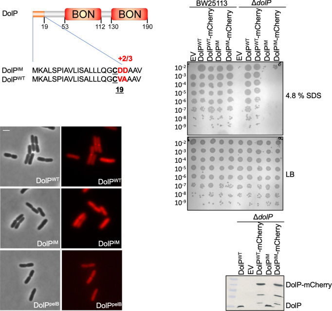
Figure 1—figure supplement 5. Localisation of DolP to the OM is required for function.

Figure 1—figure supplement 5.

The signal sequence and domain architecture of DolP are shown. The sequence changes to pET17b-dolPWT to create the construct targeting DolP to the IM (pET17b-dolPIM) are shown in red. The signal sequence of dolP was also swapped for that of pelB in order to create the construct pET17b-dolPpelB in order to target DolP to the periplasmic space with no modification. Fluorescence microscopy of ΔdolP cells expressing either DolPWT-mCherry or DolPIM-mCherry or DolPpelB-mCherry from the pET17b plasmid after growth to mid-exponential phase (OD600 ~0.4–0.8). Scale bars represent 2 μM and both phase contrast and the mCherry channel are shown in greyscale and red, respectively. The capacity of DolPWT, DolPIM, DolPWT-mCherry or DolPIM-mCherry to complement the ΔdolP mutant sensitivity phenotype was screened by dilution assay on 4.8% SDS. The expression of each construct was checked by Western blotting of total protein extracts with anti-DolP antiserum.
Figure 1—figure supplement 5—source data 1. The influence of signal sequences on DolP localisation.