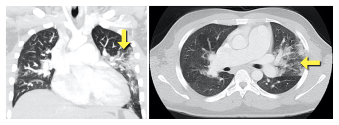
Figure 3.
CT scan showing multiple areas of parenchymal ground glass hyperdensity (yellow arrow) in the upper left lung lobe, in the middle lobe and in the lower lung lobes compatible with viral pneumonia (COVID-19), no endoluminal filling defects in the major pulmonary arterial branches, and enlarged right cardiac chambers

