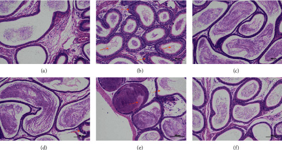
Figure 11.

Pathological observation of epididymis. Epididymis tissues obtained at the end of the experiment were stained with hematoxylin and eosin (H&E, 100x magnification): (a) NG; (b) MG; (c) HDG; (d) MDG; (e) LDG; (f) PG. NG: normal group, MG: model group, HDG: high dose group, MDG: middle dose group, LDG: low dose group, PG: positive group. Scale bar = 200 μm.
