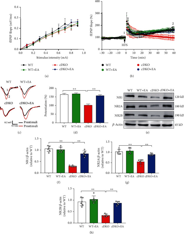
Figure 2.

EA treatment improves impaired LTP at SC-CA1 synapses and level of NMDA receptors in the hippocampus of PS cDKO mice. (a) Quantitative data of I/O curves obtained at different stimulus intensities. (b) Normalized fEPSP slopes in SC-CA1 synapses from hippocampal slices of indicated mice. (c) Representative fEPSP traces taken before (1, black lines) and 60 min after tetanus stimulation (2, red lines) from each group. Scale bar: 0.5 mV, 10 ms. (d) Quantitative data of potentiation at 60 min after tetanus stimulation (n = 7 slices from 5 mice for each group). (e) Representative Western blot of synapse-associated proteins in the hippocampus. (f)–(h) Quantification of Western blot of the synapse-associated proteins in the hippocampus (n = 6 mice for each group). The levels of synapse-associated proteins were standardized based on the respective level of β-actin. The values were expressed as relative changes to the respective WT mice, which was set to 1. Data are the mean ± S.E.M., ∗p < 0.05, ∗∗p < 0.01.
