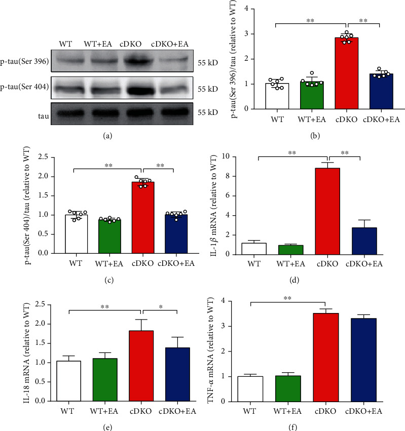
Figure 3.

EA treatment inhibits tau hyperphosphorylation and elevated inflammatory response in the hippocampus of PS cDKO mice. (a) Representative Western blot of phosphor-tau (Ser396/Ser404) in the hippocampus. (b, c) Quantification of Western blot for p-tau in the hippocampus (n = 6 mice for each group). The protein levels of p-tau were normalized with the levels of their respective total tau. The values were expressed as relative changes to the respective WT mice, which was set to 1. (d)–(f) Quantitative mRNA levels of IL-1β, IL-18, and TNF-α in the hippocampus by qRT-PCR (n = 5 mice for each group). The mRNA levels were standardized based on the respective level of β-actin. Values were expressed as relative changes to WT mice, which was set to 1. Data are the mean ± S.E.M., ∗p < 0.05, ∗∗p < 0.01.
