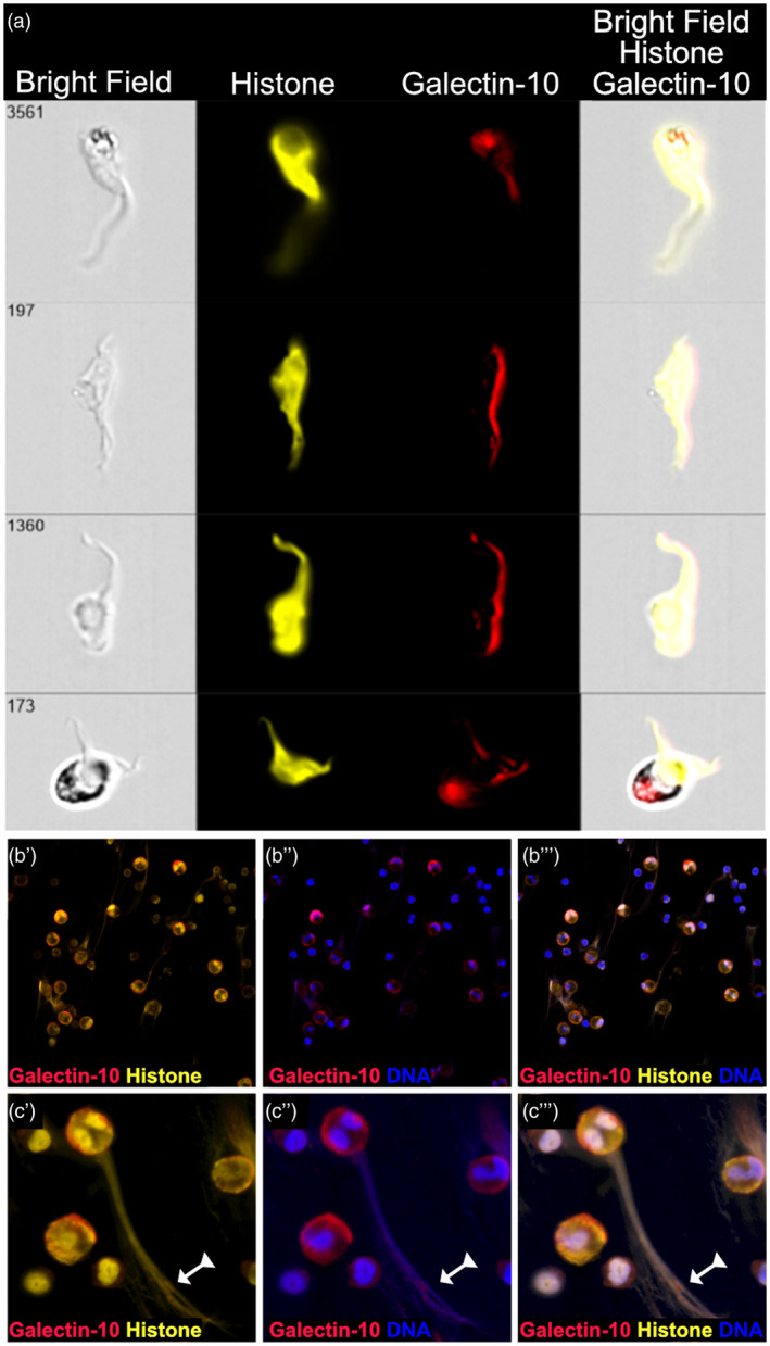
Fig. 6.

Eosinophil extracellular traps (EET) evoked by proliferating T cells contain nuclear DNA. (a) Four representative imaging flow cytometry pictures of EETs generated by eosinophils co‐cultured with CD3/CD28‐activated peripheral blood mononuclear cells (PBMC) for 1 day are shown. The EETs were stained for histones (yellow), which are associated with nuclear but not mitochondrial DNA, and for galectin‐10 (red). Bright‐field and overlay images are shown. (b) Confocal microscopy images at ×40 magnification show that EETs composed of histone (yellow) and DNA (blue) are saturated with galectin‐10 (red). (c) A large magnification of an eosinophil that has released an EET composed of histones, DNA and galectin‐10 (arrow with triangle tail). Panels labelled by a single prime (′) show staining for galectin‐10 and histone, figures labelled by a double prime (′′) show staining for galectin‐10 and DNA and those labelled with a tripple prime (′′′) show staining for galectin‐10, histone and DNA.
