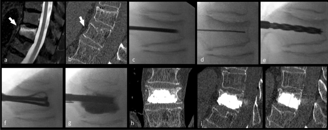
Fig. 2.
In this figure, the case of a 63-year-old female subject presented with acute back pain of 7 on the VAS. Panel a shows sagittal fat-saturation T2 weighted image and shows an A1.1 Magerl-type fracture at L2 (white arrow); Fig. 1b shows corresponding sagittal bone window CT image. In the panel c–g, the different phases of the Tektona bone remodeling procedure are given. In the panel c–g, the different phases of the Tektona bone remodeling procedure are given: c introduction of the trocar needle into the vertebral body; d the Kirschner wire has been introduced and the trocar needle removed; e the drill has been advanced over the Kirschner wire; f insertion of the VFR device and expansion of the lamella; g after the VFR device has been totally removed, the bone fillers were introduced and the bone cement has been injected. In the panel h and i, the postoperatively acquired CT is given, whereas in the panel l, the 12-month follow-up

