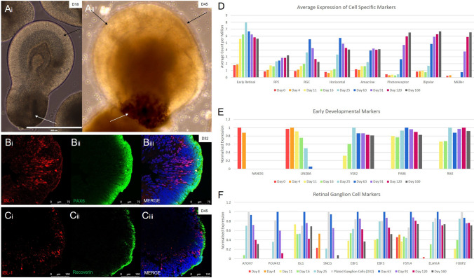
Figure 3.
Confirmation of retinal organoid development. (Ai-ii) Whole organoids were observed to have clear portions of neural retina (black arrows) and pigmented cells (white arrows) as early as day 18 (Scale Bar = 400 µm). (Bi-iii) Cross sectioned IHC analysis showed the formation of a distinct ganglion cell layer, shown here using the ganglion marker ISL1 (in red). (Ci-iii) A separation of the inner ganglion layer (shown by ISL-1 in red), and the outer photoreceptor layer (shown by the common photoreceptor progenitor marker RCVRN, in green), was seen by day 45. (D–F) Quantitative RNA sequencing data of cell specific markers showed sequential development of retinal cell types in our organoids in line with other published protocols. (D) An average of multiple markers for each cell type show cell development over time. (E) Common pluripotent and early developmental markers showed expression with comparable results to previously published protocols (Table 1). (F) Retinal ganglion cell markers show peak expression between Day 32 and 63 before decreasing over time. Ganglion subtype markers were also shown to be present (FOXP2, FSTL4), as well as markers strongly associated with ISL1 and SNCG (EBF1, EBF3).

