Figure 5.
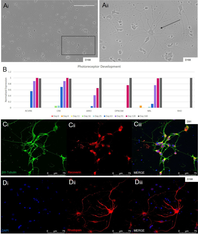
Generation of photoreceptors. (Ai-ii) Widefield images of plated photoreceptors at D168 (black arrow), obtained from dissociated organoids, show outgrowth that is morphologically different to plated ganglion cells (Shown in Fig. 6a) (Scale bar = 200 µm). (B) Quantitative RNA sequencing data of common photoreceptor progenitor markers CRX and RCVRN showed increasing expression after day 63, while the mature cone and rod markers ARR3, OPN1SW and NRL, RHO also increased over time, although not as dramatically. (Ci-iii) Expression of photoreceptor progenitor markers was seen with IHC staining of RCVRN (in red) with the general neuronal marker β-III Tubulin (in green). (Di-iii) More mature photoreceptors were stained using anti-rhodopsin antibody ROD (in red).

