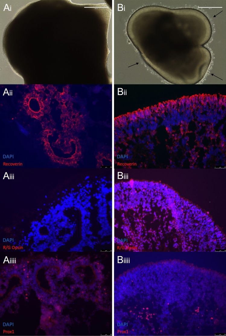
Figure 6.
Long term lamination of retinal organoids. Organoids were either ‘treated’ by the addition of the external factors FBS, Taurine, RA or T3, or left ‘untreated’ in standard RDM. A comparison between long term cultures of untreated (Ai–Aiii) and treated (Bi–Biii) retinal organoids showed significant differences. (Ai) Untreated organoids appear extremely dark and dense, and contain photoreceptor rosettes. (Bi) Treated organoids retain their golden outer layer, and develop photoreceptors outer segments (black arrows) (Scale bars = 400 µm). (Aii) Photoreceptors develop within defined rosettes on the outermost part of the untreated organoid, shown by staining with the photoreceptor progenitor marker, RCVRN (in red). (Bii) Treated organoids contain an outer layer of photoreceptors surrounding the organoid, shown by RCVRN (in red). (Aiii) Untreated organoids also do not develop Red-Green cones by Day 160, suggested by lack of RNA expression and IHC staining. (Biii) The addition of T3 in the treated organoid cultures stimulated the development of Red-Green cone photoreceptors. (Aiiii) Although having lost the lamination of the organoid and developing rosettes, untreated organoids did still develop other cells around the rosettes, such as horizontal cells, found at the bottom of the photoreceptors. (Biiii) Treated organoids retain an multi-layered appearance, with photoreceptors on the outer edge of the organoid, and other cells positioned below as found in the in vivo retina.

