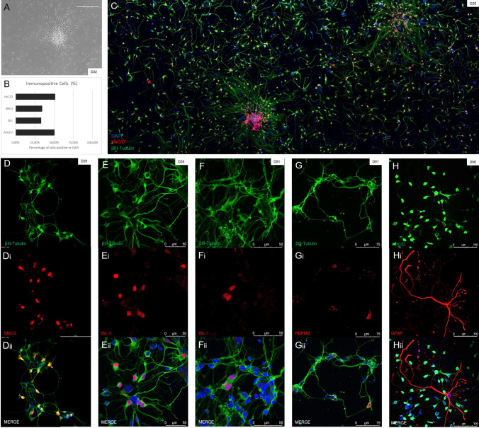
Figure 7.
Confirmation of the presence of retinal ganglion cells. (A) Widefield images of ganglion cells observed in culture showed polar cell bodies and an outgrowth of long axons by 32 days of culture. (B) When calculating the efficiency of ganglion cell generation, we found approximately 30–50% of cells were positive for ganglion cell specific markers when compared to general DAPI staining. The early ganglion marker ATOH7 showed positive staining in over 50% of cells (n = 510), with SNCG, a marker highly present in mature RGCs alongside BRN3B, showing positive staining in approximately a third of cells (n = 1779). Other commonly used RGC markers, although not completely specific, showed similar levels of positive staining. ISL1 was present in approximately a third of total cells (n = 1251), with HuC/D staining observed in almost half of all cells (n = 746). (C) After dissociating organoids at day 23, we observed a consistent amount of RGC outgrowth after 5 days. (Di-iii, Ei-iii) IHC staining of early RGC cultures using multiple ganglion markers SNCG and ISL-1 (both in red) showed positive expression and neurite outgrowth within 4 weeks of differentiation, shown by βIII-Tubulin (in green). (Fi-iii, Gi-iii) Plated cells from timepoints after 90 days of culturing show expression of the ganglion specific markers ISL-1 and RBPMS (both in red) co-stained with the general nerual marker βIII-tubulin (in green). (Hi-iii): Later stage cultures at day 96 showed expression of the RGC and amacrine marker HuC/D (in green) alongside Müller cells, identified using GFAP (in red).

