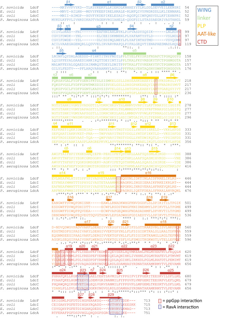
Figure 4.
Alignment of F. novicida LdcF, E. coli LdcI and LdcC, and P. aeruginosa LdcA using Clustal Omega. Partially and fully conserved residues are annotated with ‘:’ and ‘*’ respectively. Domains are coloured according to a rainbow scheme (WING domain: blue, linker: green, PLP-binding domain: yellow, AAT-like domain: orange, C-terminal domain: red), and secondary structure elements are annotated. ppGpp and RavA-interaction sites are highlighted using red and blue transparent boxes respectively.

