
Figure 5.
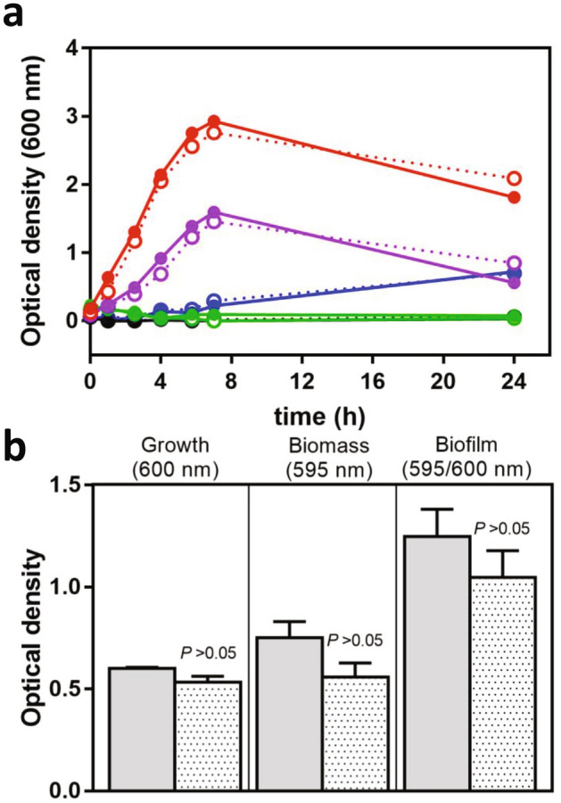
Growth and biofilm formation of F. novicida. (a) F. novicida WT (solid lines) and ΔldcF (dotted lines) were grown under shaking at 37 °C in MMH adjusted at pH 2.5 (green), pH 4 (blue), pH 6.6 (red), pH 8 (purple) or pH 10 (black) and the bacterial growth was monitored by OD600nm measurement. Results are representative of three independent trials. (b) F. novicida WT (grey columns) and ΔldcF (dotted columns) were grown for 24 h under static conditions at 37 °C in a 96-wells plates. The bacterial growth was evaluated by measurement of OD600nm and the biofilm biomass was further determined by OD595nm after Crystal violet staining. This graph corresponds to mean ± s.e.m. of three independent experiments, with at least 4 technical replicates each.