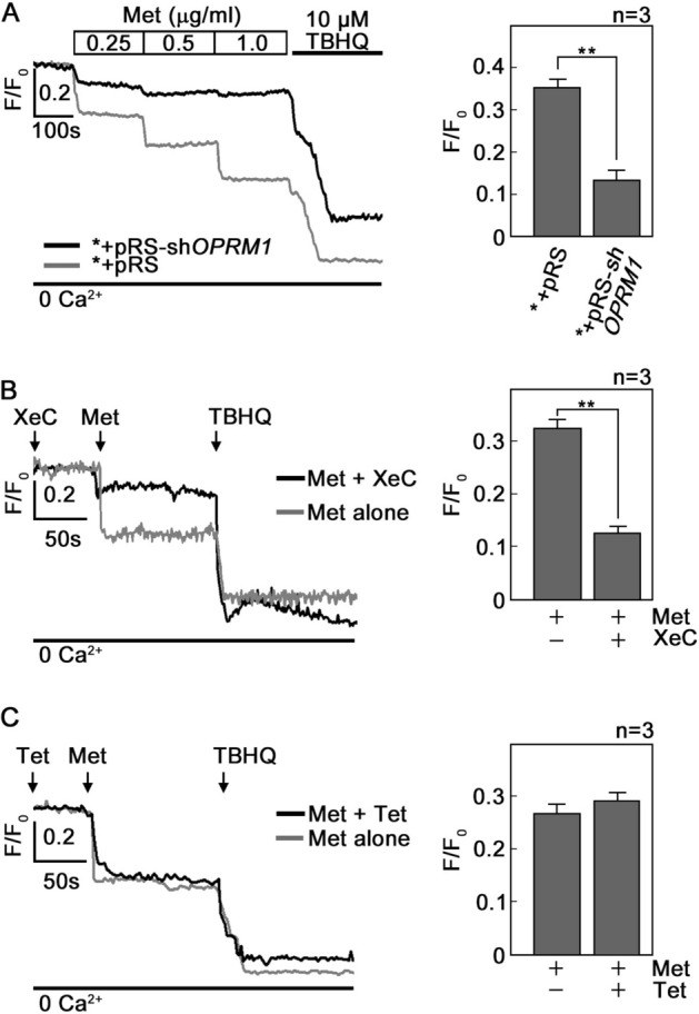
Figure 3.

d,l-Methadone induces IP3R-mediated ER Ca2+ release that is inhibited by OPRM1 loss. (A) d,l-methadone induces ER Ca2+ release that is inhibited in OPRM1-depleted cells. *+pRS and *+pRS-shOPRM1 cells were loaded with Mag-Fluo-4 AM for 30 min then treated with increasing concentrations of d,l-methadone (0.25, 0.5 and 1 μg/ml) then analyzed for ER Ca2+ release by Ca2+ imaging. The chart on the right panel shows ER Ca2+ release upon treatment of cells with 0.5 μg/ml d,l-methadone. (B) and (C). d,l-Methadone-evoked ER Ca2+ release occurs via the IP3R channel. Thirty min after loading with Mag-Fluo-4 AM, *+pRS cells were treated with xestospongin C (XeC, in B) or tetracaine (Tet in C) for 10 min. ER Ca2+ release upon d,l-methadone addition was then measured every 2 s. The right panels show ER Ca2+ release upon treatment with d,l-methadone with or without prior treatment with XeC (B) or Tet (C). Representative data from one of three independent experiments (n = 3) showing similar results. Ca2+ response after TBHQ addition indicates that cells were viable during the assay. Values are means ± SEM from three independent experiments (n = 3). **p < 0.02.
