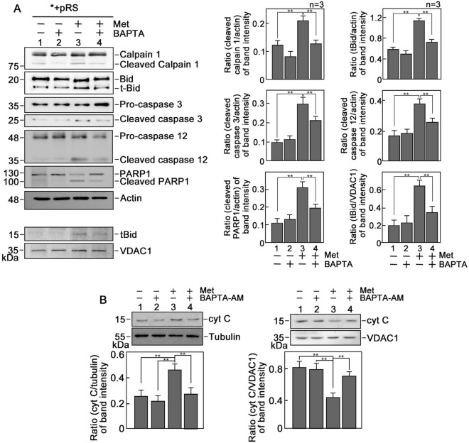
Figure 6.
d,l-Methadone induces apoptosis through upregulation of the Ca2+-mediated calpain-1-Bid-cytochrome C-caspase-3/12 apoptotic pathway. (A) Lysates of cells treated with d,l-methadone in the presence or absence of BAPTA-AM for 24 h were analyzed by SDS-PAGE and immunoblotting for calpain 1, Bid, caspase-3, cleaved caspase-3, caspase-12, PARP1 and actin. Actin blot was used as loading control. Mitochondrial fractions from cells treated as described above were also analyzed by SDS-PAGE and immunoblotting for t-Bid and VDAC1. Representative blots are from one of three independent experiments (n = 3) showing similar results. Charts on the right panel show the ratios of the levels of apoptosis-associated proteins vs actin or VDAC1, with the actin and VDAC1 values normalized to 1.0. The charts correspond to the densitometry analysis of the representative blots shown on the left panel. (B) d,l-Methadone increases cytochrome C (cyt C) level in the cytosol. Cytosolic and mitochondrial fractions from cells pre-treated or untreated with 0.5 μM BAPTA-AM for 30 min then treated or untreated with 0.5 μg/ml d,l-methadone for 12 h were analyzed by SDS-PAGE and immunoblotting for cyt C, tubulin and VDAC1 (upper panels). Tubulin and VDAC1 were used as loading controls for cytosolic and mitochondrial fractions, respectively. Representative blots are from one of three independent experiments showing similar results. The lower panels show the ratios of cyt C vs tubulin or VDAC1 levels, with tubulin or VDAC1 levels normalized to 1.0. Values are means ± SEM of the three independent experiments (n = 3). **p < 0.05.

