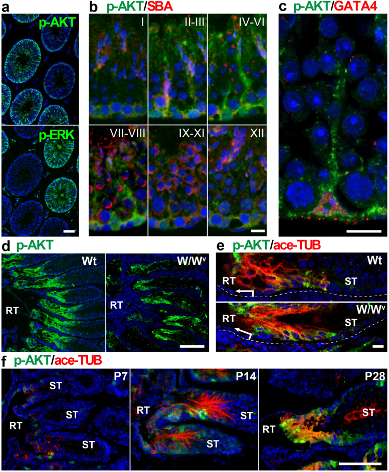
Figure 1.
High phosphorylated AKT (p-AKT) signals in the SV epithelia. (a–c) Anti-p-AKT (green in a–c), anti-phosphorylated ERK (p-ERK; green in a), SBA lectin (acrosome staining; red in b), and anti-GATA4 (red in c) staining of the convoluted ST of adult wild-type mouse testes. In plate (a), anti-p-AKT and p-ERK immunostaining of two serial sections shows reversed complementary expression patterns of p-AKT and p-ERK in the ST. In plate (c), p-AKT signals were observed mainly in the cytoplasmic region of GATA4-positive Sertoli cells. In plate (b), roman numerals indicate the seminiferous epithelium cycle stage. (d,e) Anti-p-AKT (green) and anti-acetylated tubulin (ace-TUB; red) double staining of the proximal region of adult wild-type (Wt) and W/Wv testes, showing the transient region of RT and ST. Plate (e) shows the SV regions in plate (d) at higher magnifications. (f) Anti-p-AKT (green) and anti-ace-TUB (red) double staining of the proximal ST in the wild-type testes at postnatal (P) days 7, 14, and 28. RT, rete testis. ST, convoluted seminiferous tubules. Scale bars represent 50 μm in (a), 10 μm in (b,c), 200 μm in (d), 20 μm in (e), 100 μm in (f).

