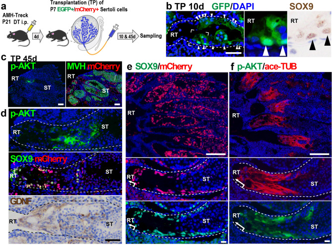
Figure 2.
Regeneration of the SV epithelia by transplanted Sertoli cells adjacent to the RT. (a) Schematic representation of Sertoli cell ablation/transplantation experiment. GFP or mCherry-positive Sertoli cells (P7) were injected into the empty STs of recipient AMH-Treck males that were pretreated with DT at P21 (P21) to ablate endogenous Sertoli cells (n = 4). (b) Donor-derived (cytoplasmic/nuclear GFP-positive) Sertoli cells (arrowheads) were settled in the presumptive SV region adjacent to the host-derived (GFP-negative) RT in the recipient Tg male at day 10 post-transplantation. Insets, magnifications of two serial sections stained with anti-GFP (green) and anti-SOX9 (brown) antibodies respectively. (c–f) Anti-p-AKT, anti-SOX9, anti-MVH (germ cell marker), or anti-GDNF staining, together with anti-mCherry staining, in the proximal region of the Tg testes at day 45 post-transplantation. The donor-derived Sertoli (mCherry/SOX9-double positive) cells regenerated SV-like epithelia positive for anti-ace-TUB, anti-p-AKT, and anti-GDNF staining between the RT and the convoluted ST with several patches of active spermatogenesis (ST; MVH-positive in c; meiotic and post-meiotic germ cells in hematoxylin- and 4′,6-diamidino-2-phenylindole (DAPI)-stained images in d–f). Two panels of c, upper two panels of d, and two top and four lower panels of (e,f) represent serial sections. Broken lines are the outlines of the ST. DT diphtheria toxin, RT rete testis, ST seminiferous tubule. Scale bars represent 50 μm in (b,d), 100 μm in (c), 200 μm in (e,f) (upper), 20 μm in (e,f) (lower).

