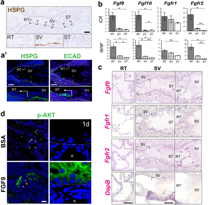
Figure 3.
Expression profiles of FGF-associated genes and their association with p-AKT expression in the SV epithelia. (a) HSPG immunohistochemistry (brown in a, green in the left plate of a’), showing high HSPG expression in the basement membrane of the SV region, compared to those in the convoluted ST and the RT marked by ECAD (E-cadherin; Cadherin-1) immunohistochemistry (green in the right plate of a’). (b) Quantitative RT-PCR analysis of FGF-related genes (Fgf9, Fgf10, Fgfr1 and Fgfr2) in the RT, SV, and convoluted ST fragments from wild-type ICR mouse testis (n = 4) and W/Wv testes (n = 5). Analysis was performed using a paired t-test. *p < 0.05; **p < 0.01. Broken horizontal line, expression level in the wild-type convoluted ST with active spermatogenesis. (c) In situ hybridization images of wild-type mouse testis, showing the expression of Fgf9 in the RT, and expressions of Fgfr1 and Fgfr2 in both the RT and the SV. DapB was used as a negative control probe, showing little non-specific staining in the RT and the SV region. (d) Anti-p-AKT (green) staining of W/Wv mutant mouse testis at 24 h after FGF9-soaked bead transplantation, showing an ectopic appearance of p-AKT signals in the ST close to the transplanted FGF9-soaked bead (asterisk), not in the tubules far away from the bead or near BSA-soaked beads (asterisk). The right-most panel shows the magnified image of the p-AKT-positive Sertoli cells indicated by the broken square (broken line, the tubular wall). Figures in b were generated by using R60. Asterisk beads, RT rete testis, SV Sertoli valve, ST seminiferous tubule. Scale bars represent 50 µm.

