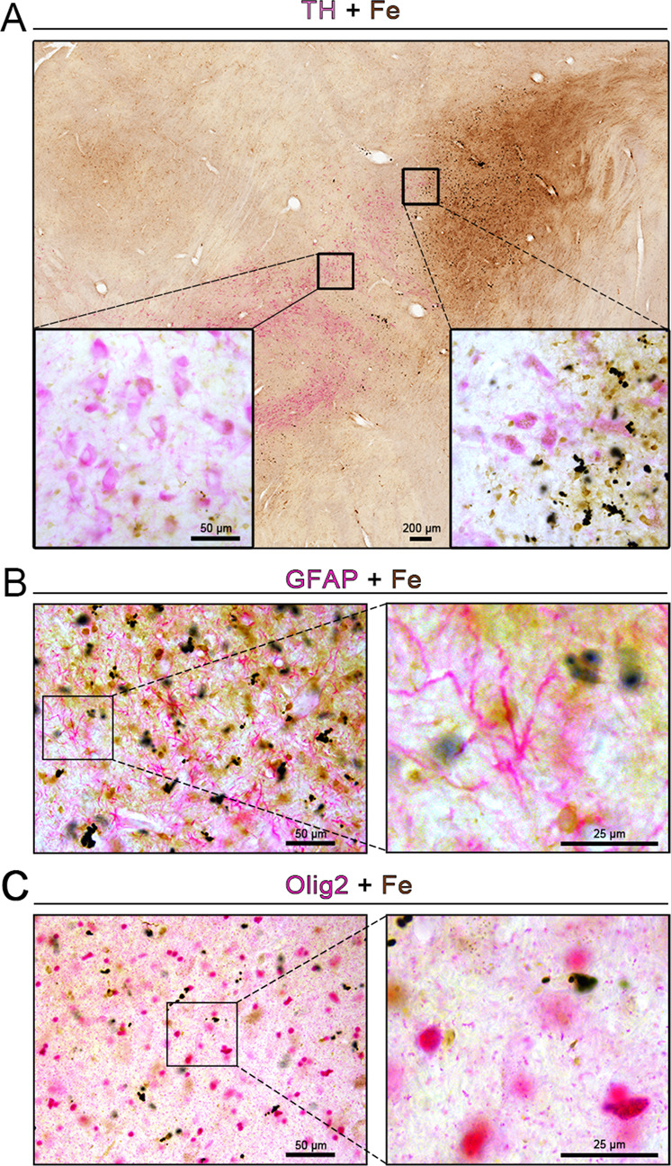
Fig. 4. Cellular localization of iron deposits in the substantia nigra.

A–C Double-labeling of chemically stained iron ions (dark brown) along with immunohistochemically stained with TH (A), GFAP (B), or Olig2 (C) antibody (magenta) showed heavy iron deposition 17 months after treatment with α-syn PFFs. The inset images depict the enlarged small boxes in A–C. Scale bars: 200 μm in A and 50 µm in the inset; 50 µm in B, C, and 25 µm in the enlarged images.
