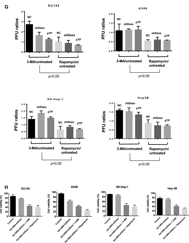
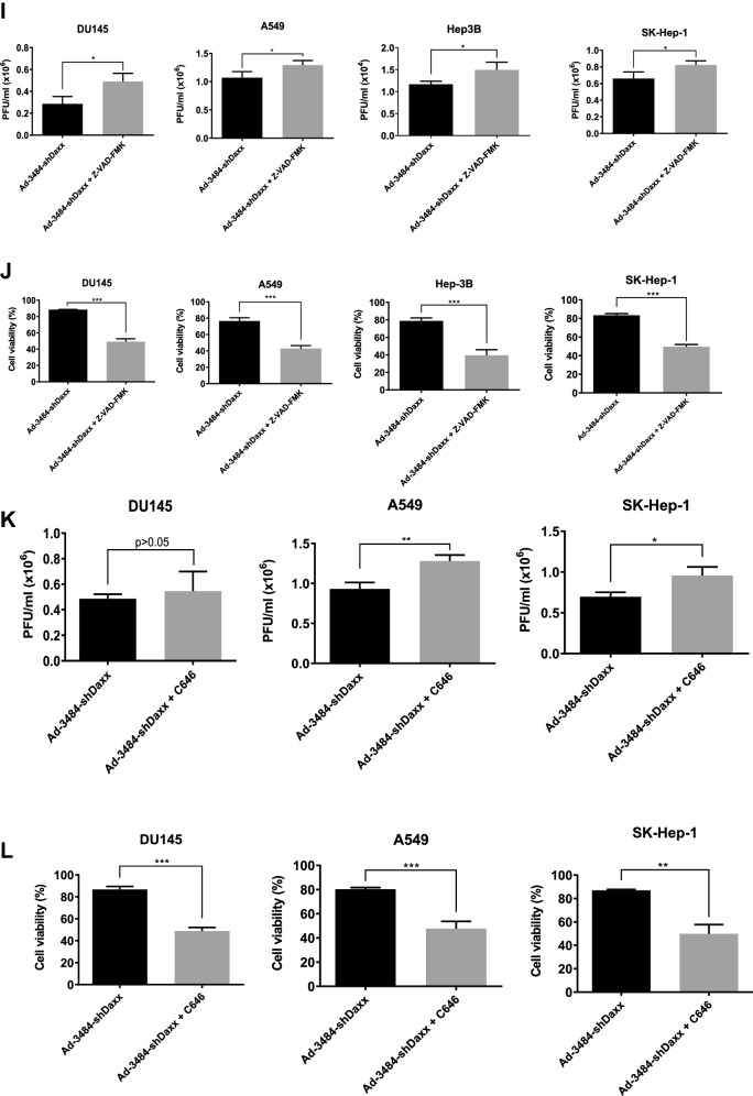
Figure 4.
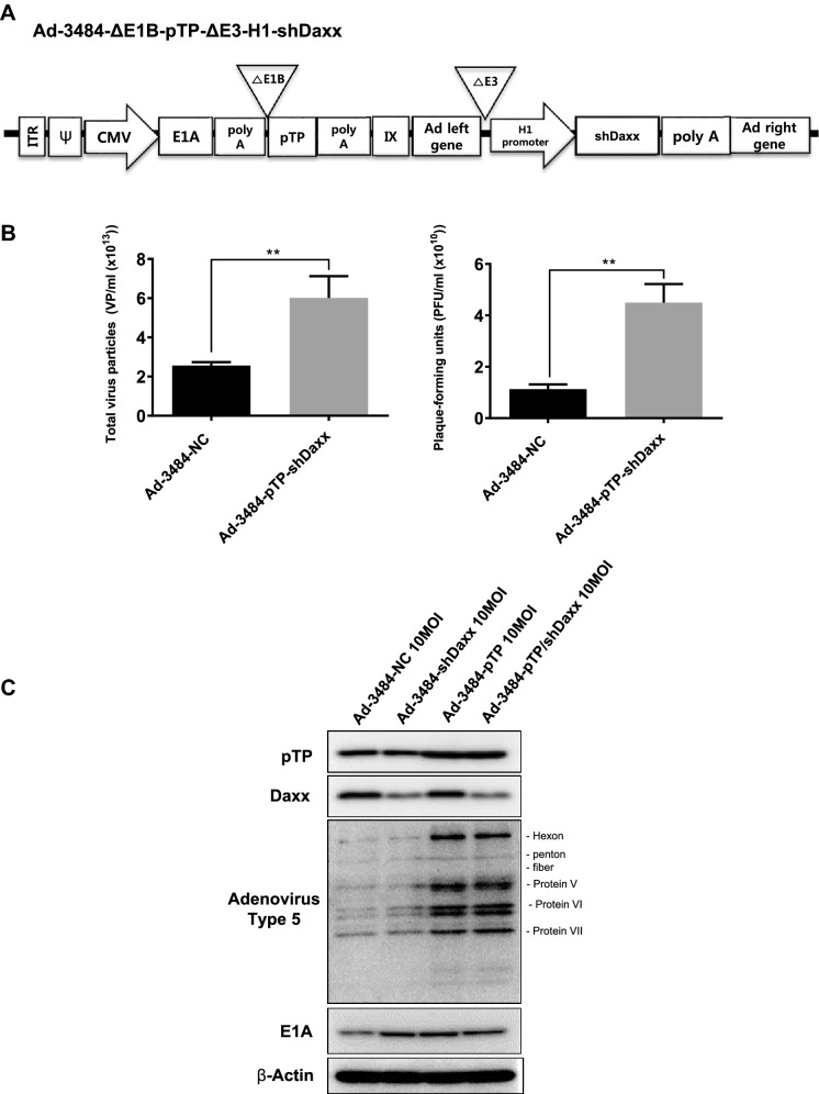
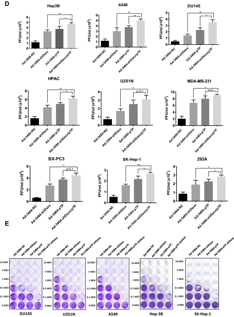
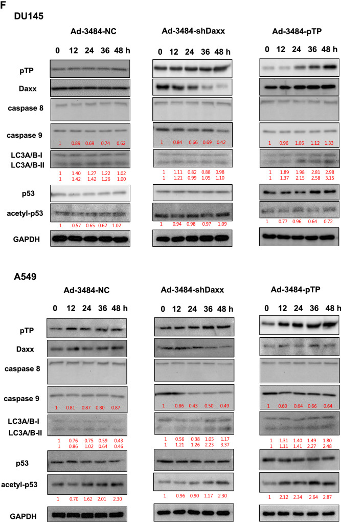
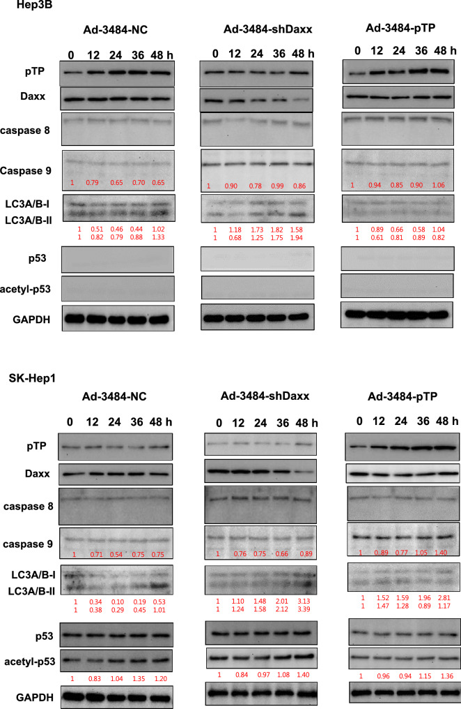
Enhanced virus production by both pTP and Daxx shRNA expressing E1B55K-deleted oncolytic adenovirus (Ad-3484-pTP-shDaxx). (A) Scheme of pTP and Daxx shRNA-both expressing E1B55K-deleted oncolytic adenovirus. (B) Virus particle amounts were determined after purification through a CsCl gradient and Tris-dialysis of virus soup from 293A cells. Following infection with the same amount of infectious oncolytic adenovirus (Ad-3484-NC and Ad-3484-pTP-shDaxx), total virus particles (VP/ml) (left) and plaque-forming units (PFU/ml) (right) were calculated. Error bars represent standard errors from three independent experiments. P values less than 0.05 were considered statistically significant (**P < 0.01). (C) Western blot analysis for the detection of protein levels, including adenovirus type 5, from DU145 cells infected with 10 MOI of Ad-3484-NC, Ad-3484-shDaxx, Ad-3484-pTP, or Ad-3484-pTP-shDaxx for 48 h. (D) PFU titration assays of adenovirus produced by each human cancer cell line following Ad-3484-NC, Ad-3484-shDaxx, Ad-3484-pTP, or Ad-3484-pTP-shDaxx infections at MOI 50 for 4 h, and incubation for 48 h with media exchanged after PBS washing. Adenovirus packaging cells (293A) were infected at MOI 0.1. Error bars represent standard errors from three independent experiments. (E) Oncolytic assays of various cancer cells (DU145, U251N, A549, Hep3B, and SK-Hep1) infected with Ad-3484-NC, Ad-3484-shDaxx, Ad-3484-pTP, or Ad-3484-pTP-shDaxx at MOIs between 0 and 10, for 72 h. All cells remaining on the plates were fixed with 4% paraformaldehyde and stained with 0.05% crystal violet. (F) Western blot analysis for detection of caspase 8, 9, LC3A/B-I, II, p53 and acetylated p53 (Lys382) protein levels in various cancer cells infected with 10 MOIs of Ad-3484-NC or Ad-3484-shDaxx or Ad-3484-pTP for each indicated time. The numbers indicate the relative band intensity of infected samples for each indicated time to control (0 h) after band intensities were measured with a densitometer. (G) Plaque-forming unit (PFU) titration assays of adenovirus in various cancer cells treated with autophagy inhibitor (3-MA 10 mM) or autophagy enhancer (rapamycin 50 nM) for 44 h after 4 h of infections with 10 MOIs of Ad-3484-NC, Ad-3484-shDaxx, or Ad-3484-pTP. PFU ratios mean that viral titer of 3-MA or rapamycin-treated cancer cells after infection NC, shDaxx or pTP virus divided by viral titer of untreated cancer cells after infection NC, shDaxx or pTP virus respectively. Error bars represent standard errors from three independent experiments. NC, Ad-3484-NC; shDaxx, Ad-3484-shDaxx; pTP, Ad-3484-pTP (H) Cell viability in various cancer cells treated with autophagy inhibitor (3-MA 10 mM) or autophagy enhancer (rapamycin 50 nM) for 44 h after 4 h of infections with 10 MOIs of Ad-3484-shDaxx. Cell viability was estimated by trypan blue exclusion. Error bars represent standard errors from three independent experiments. (I) Plaque-forming unit (PFU) titration assays of adenovirus in various cancer cells treated with pan caspase inhibitor (Z-VAD-FMK 10 μM) for 44 h after 4 h of infections with 10 MOIs of Ad-3484-shDaxx Error bars represent standard errors from three independent experiments. P values less than 0.05 were considered statistically significant (*P < 0.05). (J) Cell viability in various cancer cells treated with pan caspase inhibitor (Z-VAD-FMK 10 μM) for 44 h after 4 h of infections with 10 MOIs of Ad-3484-shDaxx. Cell viability was estimated by trypan blue exclusion. Error bars represent standard errors from three independent experiments. P values less than 0.05 were considered statistically significant (***P < 0.001). (K) Plaque-forming unit (PFU) titration assays of adenovirus in various cancer cells treated with histone acetyltransferase inhibitor (C646 25 μM) for 44 h after 4 h of infections with 10 MOIs of Ad-3484-shDaxx. Error bars represent standard errors from three independent experiments. P values less than 0.05 were considered statistically significant (*P < 0.05; **P < 0.01). (L) Cell viability in DU145, A549 and SK-Hep1 treated with histone acetyltransferase inhibitor (C646 25 μM) for 44 h after 4 h of infections with 10 MOIs of Ad-3484-shDaxx. Cell viability was estimated by trypan blue exclusion. Error bars represent standard errors from three independent experiments. P values less than 0.05 were considered statistically significant (***P < 0.001).

