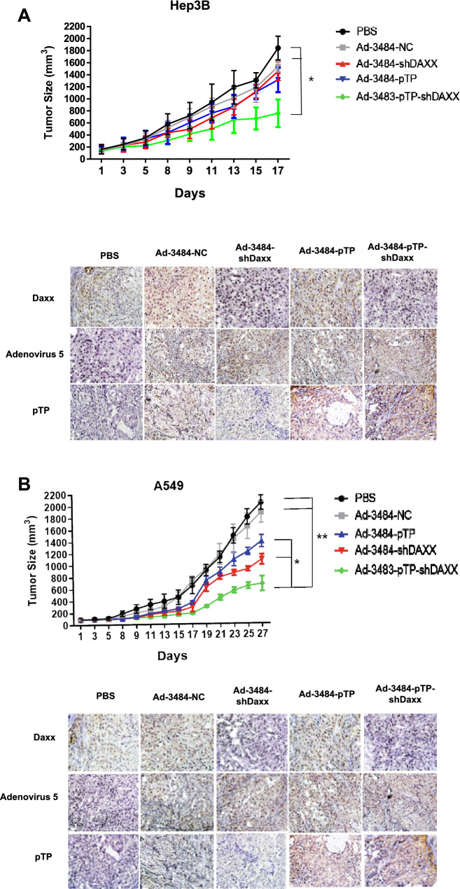
Figure 5.
Tumor size measurements and immunohistochemical images of tumors. Hep3B cells (A) or A549 cells (B) in Matrigel were injected subcutaneously in the abdominal region of BALB/c athymic nude mice, and intratumoral injections of PBS, or of each virus indicated, were repeated every other day for a total of three injections. Tumor volume was measured and calculated on the indicated days using the following formula: Volume (mm3) = 0.52 × (Length) × (Width)2 (n = 5 per group) (upper). Error bars represent standard error from five mice in each experimental group. The asterisk indicates a significant difference between each comparative group (*P < 0.05; **P < 0.01). Tumor tissues after euthanasia from each group (n = 2 per group) for immunohistochemistry were harvested 7 days following virus infections. Adenovirus 5, pTP or Daxx primary antibodies were incubated overnight at 4 °C on tissue slides. Images were obtained by light microscopy (200×). (lower).

