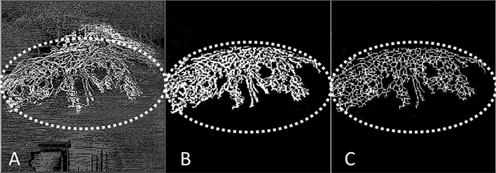
Figure 2.
Image processing technique using an in-house automated programme written in MATLAB (Mathworks, Inc., Natick, MA, USA); the region of interest (ROI) is the area demarcated by the dotted line. (A) Example of corneal vascularisation captured using anterior segment optical coherence tomography (AS-OCT) angiography (Nidek RS-3000, Tokyo, Japan); (B) the overlay image is binarized at the ROI where white pixels represented the blood vessels and black represented the background—vessel density (VD) is the area of white pixels as a percentage of the whole binarized image; (C) Skeletonization of the binarized images, where all the vessel segments were reduced to one pixel-wide segment—vessel branch area (VBA) is the total length of the vessel branch as a percentage of the whole binarized image; the VD as a percentage of the VBA, represents the vessel width of the corneal vessels in the ROI.

