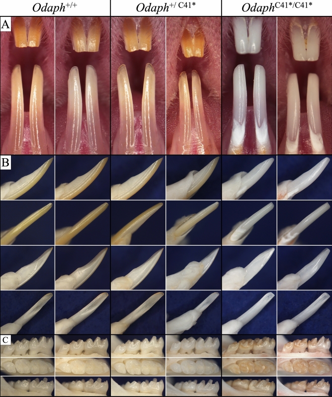
Figure 1.
Appearance of Odaph+/+, Odaph+/C41*, and OdaphC41*/C41* Incisors and Molars at 7-weeks. There were no appreciable differences in dental phenotype observed between Odaph+/+ and Odaph+/C41* mice. Tooth size and morphology were similar in the three genotypes. (A) Frontal view of maxillary and mandibular incisors. The OdaphC41*/C41* incisors appeared to be severely hypomineralized. Their incisors were chalky white and the enamel had abraded from dentin surface down to the cervical region. (B) Mesial, labial, distal, and lingual (lower right) views of the mandibular incisors. The OdaphC41*/C41* incisal edge is flat and appears to be shorter than the wild-type due to attrition. (C) Buccal, Occlusal, and Lingual views of mandibular molars. No differences in alveolar bone level were observed among the three genotypes. The OdaphC41*/C41* molars were discolored and had undergone significant attrition.

