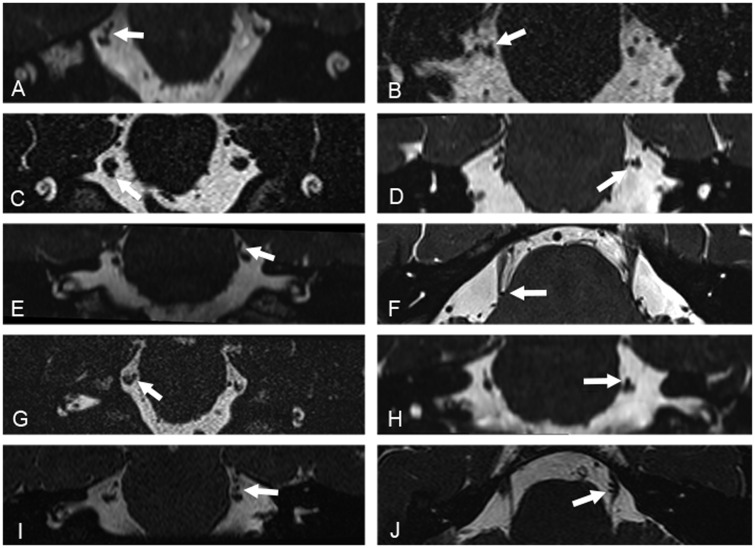
Figure 1.
High-resolution volumetric 3-D CISS and SPACE MRI acquisitions. Acquisitions were reconstructed in the coronal (A–E and G–I) and axial (F and J) planes, and demonstrate contact of a segment of the superior cerebellar artery with the cisternal segment of the trigeminal nerve (white arrows). Images in the left column show contact with no morphological changes of the corresponding segment of the trigeminal nerve. Images in the right column show contact with morphological changes, with deviation and distortion of the corresponding adjacent cisternal segment of the trigeminal nerve.

