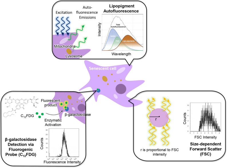
Figure 1.
Various methods used for automatic quantification of hMSCs senescence in this study. Top: New autofluorescence method that employs cell lasing through endogenous fluorophores to collect autofluorescence signals. Bottom left: fluorescence-based detection of -galactosidase activities using fluorogenic substrate CFDG through enzymatic activation and flow cytometry analysis. Bottom right: flow cytometry forward scatter (FSC) measurements for hMSCs cell size determination. All three flow cytometer and autofluorescence methods in quantification of hMSCs senescence were benchmarked with the cytochemical -galactosidase staining method.

