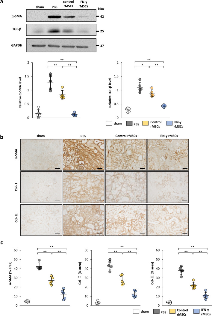
Figure 2.
Anti-fibrotic effects of IFN-γ-treated mesenchymal stem cells (MSCs) in the kidney of IRI rats. IFN-γ-treated or untreated rat MSCs were injected immediately after IRI induction. Twenty-one days later, protein levels of fibrosis markers were evaluated by western blot (a) and immunohistochemical (b, c) analyses. (a) Western blot analysis of α-SMA and TGF-β1 in the rat kidney cortex. Protein levels were normalized to GAPDH levels (n = 5 in each group). The blots are the cropped images from different parts of the same gel. Full-length gel images are provided in the supplementary file. The samples derive from the same experiment and that gels were processed in parallel. (b) Representative immunohistochemical staining of α-SMA, Collagen type I (Col-I), and Collagen type III (Col-III) in kidney sections. (scale bar, 100 µm). (c) Quantification of α-SMA-, Col-I-, and Col-III-positive areas (n = 5 in each group). Data are presented as the mean ± SD. **p < 0.01. Sham, non-IRI procedure; PBS, PBS injection; control rMSCs, rat MSCs injection; IFN-γ rMSCs, injection of IFN-γ-treated rMSCs.

