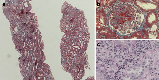
Figure 1.
Kidney biopsy results. Kidney sections under light microscopy show four glomeruli, one of which shows a cellular crescent (a: Masson’s trichrome staining, original magnification ×40). The cellular crescent has fibrinoid necrosis (b: Masson’s trichrome staining, original magnification ×400). Peritubular capillaries contain inflammatory cells (c: Periodic acid-Schiff staining, original magnification ×400).

