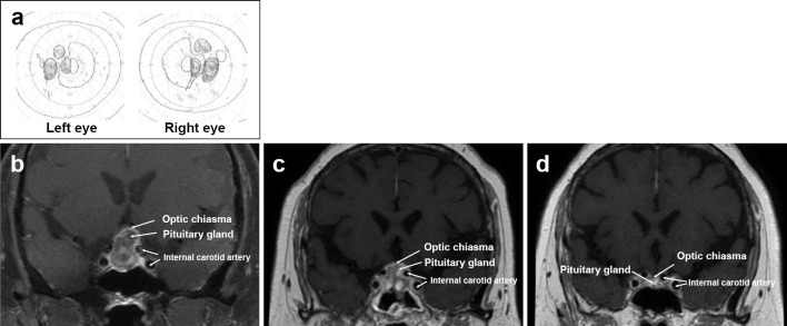
Figure 5.
MRI findings at the onset of constriction of the visual field and from one to six months after rituximab therapy. The visual field test shows bitemporal hemianopsia (a). The enlarged pituitary gland compresses the optic chiasma (b). One month after rituximab therapy, the pituitary gland is reduced in size. This leads to the amelioration of the displacement of the optic chiasma (c). Six months after rituximab therapy, follow-up MRI shows the further decrease in the pituitary gland size (d).

