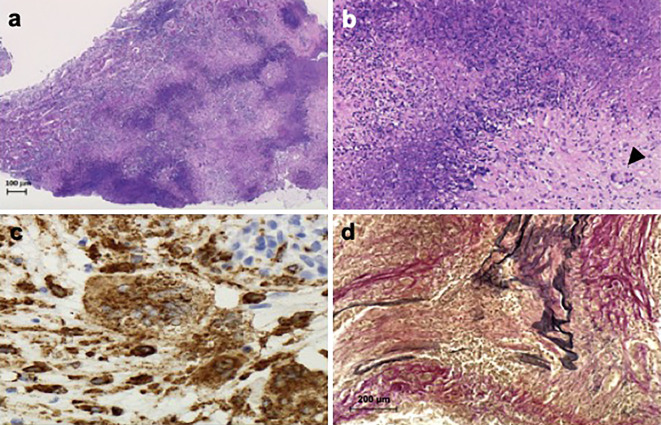
Figure 6.
Pituitary biopsy results. Pituitary sections under light microscopy show geographic necrosis [a: Hematoxylin and Eosin (H&E) staining, original magnification ×40]. Epithelioid cells and multinucleated giant cells were observed (b: H&E staining, black arrowhead shows multinucleated giant cells, original magnification ×100). These cells are CD68-positive (c: CD68 staining, original magnification ×400). Middle-sized arteries shown medial thickening and intimal proliferation. Many inflammatory cells have infiltrated into the arterial walls. The internal elastic membrane is ruptured (d: Elastica Van Gieson stain, original magnification ×200).

