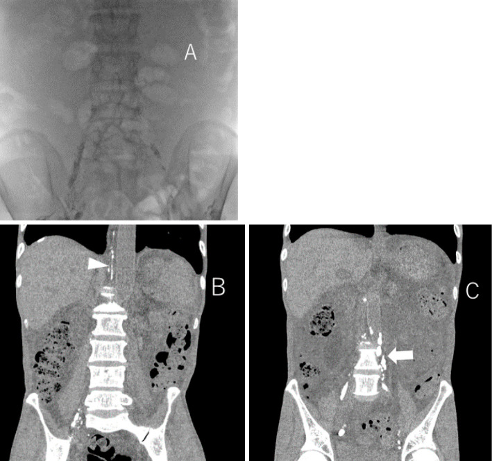
Figure 4.
Lymphangiogram was performed from the left and right inguinal lymph nodes (A) and abdominal CT was taken the day after lymphangiography. Inflow to the thoracic duct can be confirmed by lymphangiography performed from the right inguinal lymph node (arrow head) (B) but, lymphangiography performed from the left inguinal lymph node showed that the lymph vessels were occluded at the level of the 4th lumbar spine (arrow) (C).

