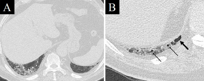
Figure 2.
Chest computed tomography images at the bottom of the lung on admission. At some distance from the diaphragm, no cystic lesions could be seen just below the pleura, such as a honeycomb lung, although GGO and reticular shadows were found (A). A cystic lesion (thick arrow) and bronchioles close to the pleura (thin arrows), suggestive of mild chronic fibrosis, were found just below the pleura near the diaphragm (B).

