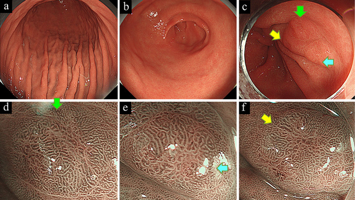
Figure 1.
White-light endoscopy showed a non-atrophic gastric mucosa of the gastric body (a) and antrum (b), suggesting an Helicobacter pylori-uninfected stomach. Three depressed lesions (arrows) were observed in the prepyloric area, showing a gastritis-like appearance (c). Narrow-band imaging with magnification endoscopy showed slightly irregularly shaped papillary or tubular microstructures that were either unclearly demarcated from the surrounding mucosa or partially covered by a non-neoplastic epithelium (d-f).

