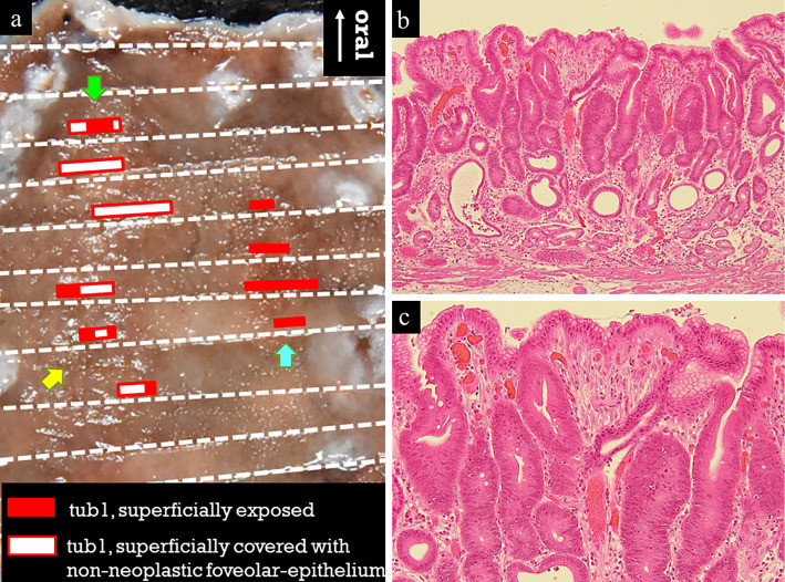
Figure 2.
Histologic mapping of the resected specimen showed three mucosal cancers. The red square indicates superficially exposed tubular adenocarcinoma, and the white square with red border indicates tubular adenocarcinoma superficially covered with a non-neoplastic foveolar-epithelium (a). In the lesion indicated by the green arrow, an irregularly shaped tubular neoplastic duct was confirmed in the middle layer of the lamina propria (b). The neoplastic cells had either roundish or polygonal nuclei lacking polarity, and the lesion surface was partially covered by a non-neoplastic foveolar epithelium (c). Only slight stromal inflammation was observed, and no spirillum was detected, thus suggesting the presence of a Helicobacter pylori-uninfected pyloric gland mucosa.

