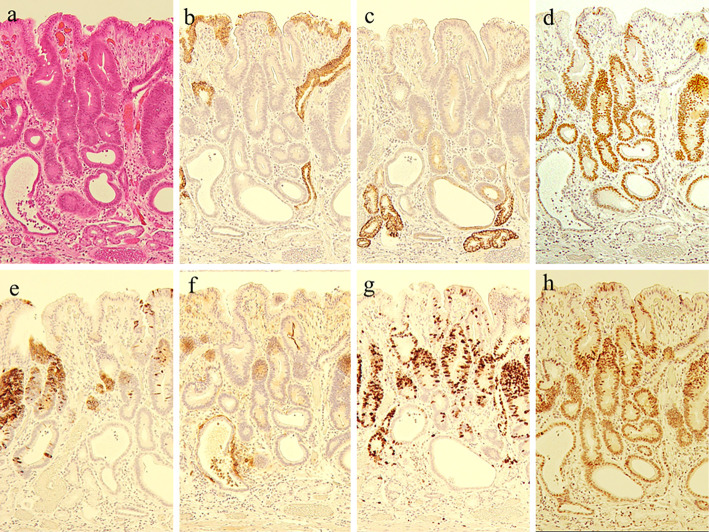
Figure 3.
a: Hematoxylin and Eosin staining, b: MUC5AC, c: MUC6, d: CDX2, e: MUC2, f: CD10, g: Ki-67, h: p53. The neoplasm expressed CDX2, MUC2, and CD10, showing immunohistochemical characteristics suggestive of an intestinal mucin phenotype. Ki-67 was highly overexpressed (labeling index, 65.2%), and p53 was diffusely highly overexpressed by neoplastic cells.

