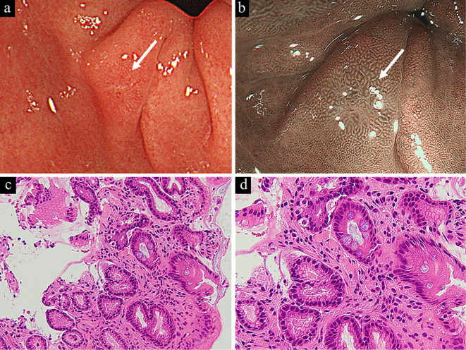
Figure 4.
A small erosive lesion was detected in the prepyloric area as indicated by the white arrow (a), and narrow-band imaging with magnification endoscopy suggested benign inflammatory findings (b). A histologic examination of a biopsy specimen showed a non-neoplastic foveolar epithelium with intestinal metaplasia (c). Goblet cells were sporadically observed, as was mild stromal inflammation with fibrosis (d).

