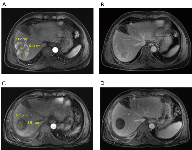
Figure 1.
Representative images of MR before TACE and after TACE. (A) The right lobe of the liver was occupied by 7.62 cm × 5.14 cm before TACE. MR artery showed obvious enhancement and rich blood supply; (B) MR portal vein stage before TACE showed rapid dissolution of contrast agent in lesions; (C) three and a half months after TACE, the lesion was significantly smaller (5.15 cm × 3.91 cm) than that before TACE. MR arterial phase showed that tumor enhancement had completely disappeared; (D) three and a half months after TACE, MR portal vein stage showed clear outline of the lesion and complete inactivation of the tumor.

