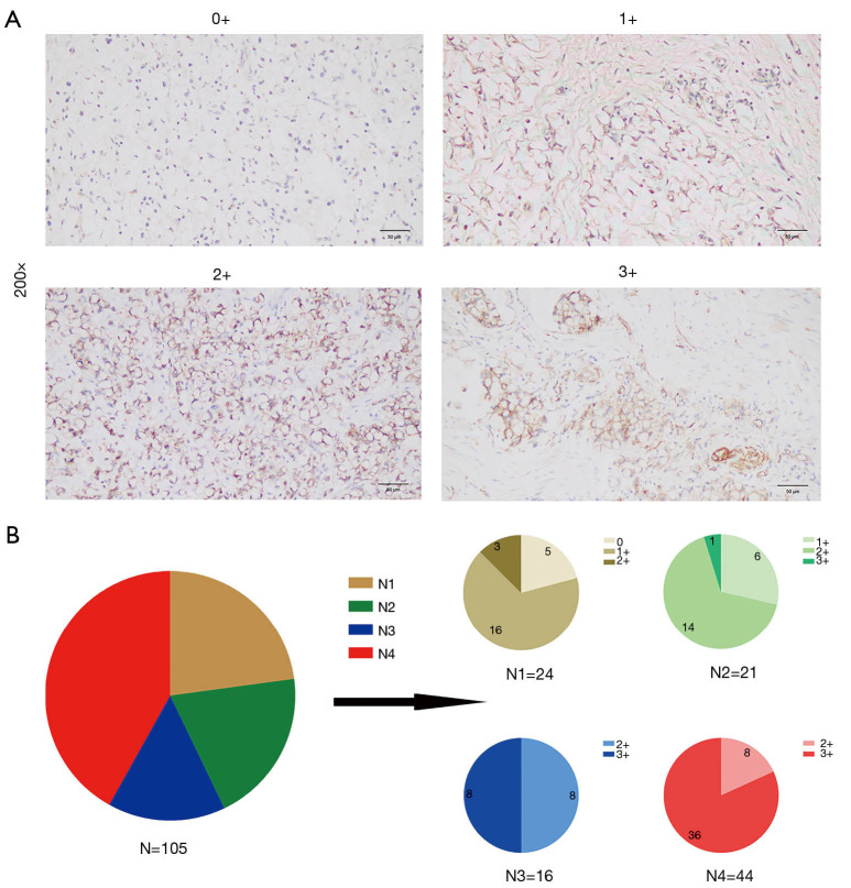
Figure 1.
Expression of CLDN18.2 in advanced gastric SRCC. (A) Micrographs of representative stained tissues: 0+, 1+, 2+ and 3+ staining intensity. The magnification was 200×. (B) The graph depicted the distribution of CLDN18.2 staining percentages and intensities in tumor cells from advanced gastric SRCC patient samples. N, total number of cases; N1, number of cases that had 0–25% CLDN18.2 positive tumor cells; N2, number of cases that had 26–50% CLDN18.2 positive tumor cells; N3, number of cases that had 51–75% CLDN18.2 positive tumor cells; N4, number of cases that had 76–100% CLDN18.2 positive tumor cells. SRCC, signet-ring cell carcinoma.

