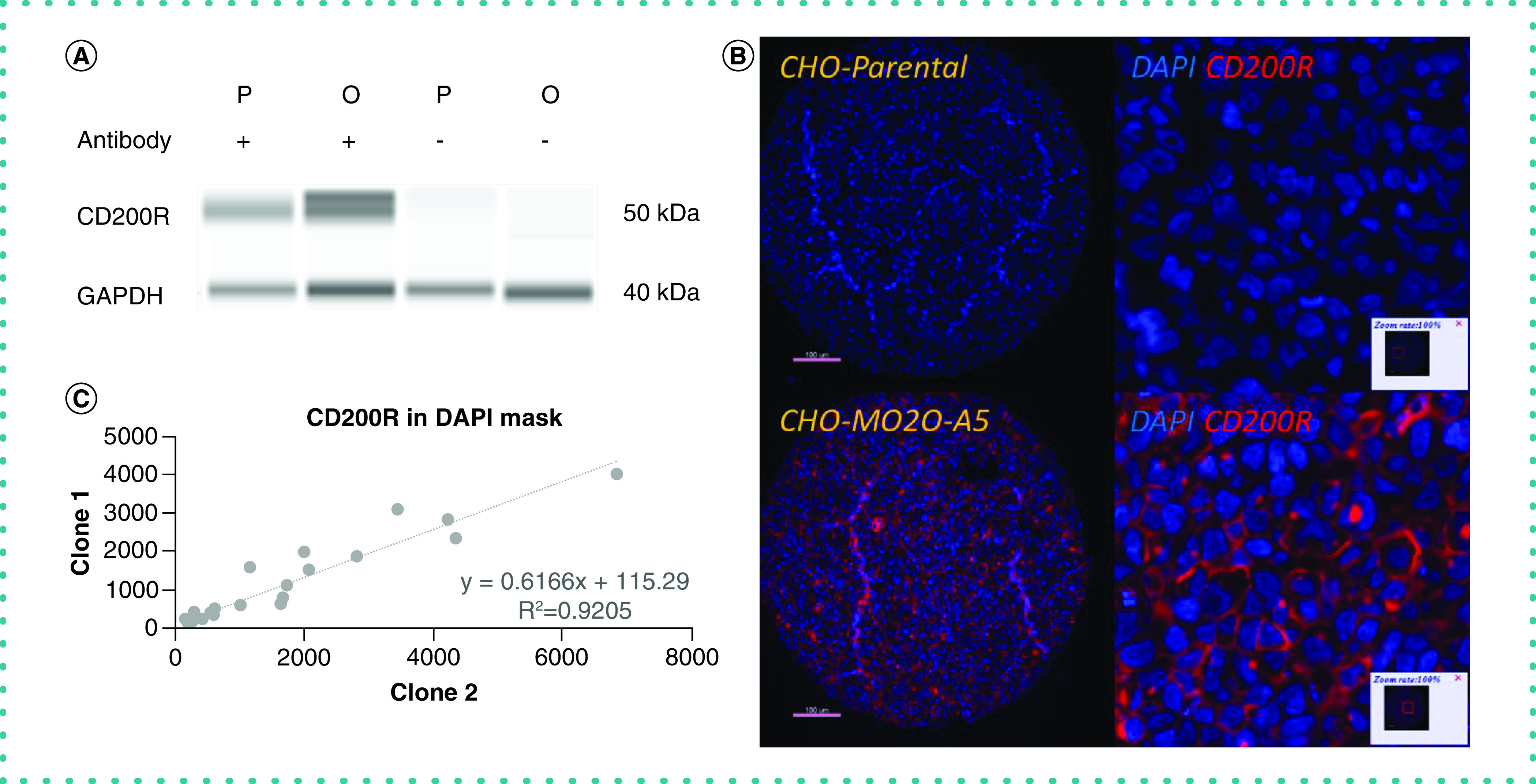
Figure 3. . Validation of CD200R antibody.

(A) Orthogonal methods of validation. Western blot exhibiting that the candidate antibody recognizes CD200R. Parental CHO cell line with anti-CD200R antibody shows weak signal (lane 1); MO2O-A5 CHO cell line with anti-CD200R antibody shows strong signal (lane 2); parental CHO cell line without antibody (lane 3) and MO2O-A5 CHO cell line without antibody (lane 4) show no signal. (B) Genetic methods of validation. CHO parental cell line has basal levels of CD200R expression and stains negative for CD200R (top); CHO MO2O-A5 cell line overexpresses CD200R and shows clear membranous staining pattern for CD200R (bottom). Isotype-specific HRP-conjugated secondary antibodies were used with a tyramide-based amplification system to generate the fluorescent signal. The Cy5 channel was used for visualization of the CD200 antibody. (C) Independent epitope validation. Scatter plot showing good correlation between two antibodies that bind to nonoverlapping epitopes of human CD200R.
CHO: Chinese hamster ovary; DAPI: 4′,6-diamidino-2-phenylindole; GAPDH: Glyceraldehyde 3-phosphate dehydrogenase; HRP: Horseradish peroxidase.
