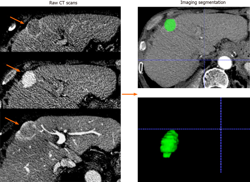
Figure 2.
Imaging segmentation. The images were acquired form a 65-year-old man with hepatocellular carcinoma (orange arrows). Serial raw computed tomography scans obtained before transarterial chemoembolization treatment; regions of interest were manually depicted along with the tumor outline on each axial slice and automatically merged into a volume of interest (green area).

