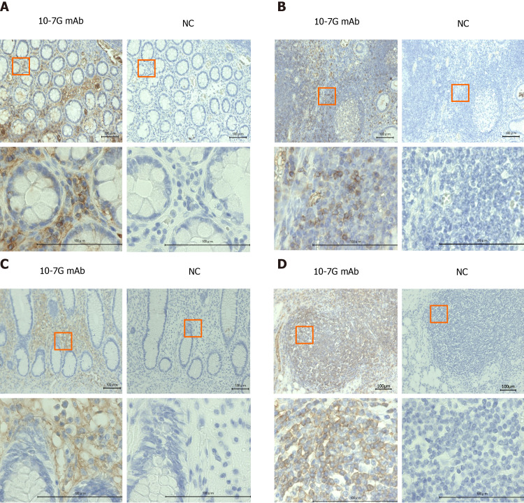
Figure 2.
Immunohistochemical study of ulcerative colitis and Crohn’s disease intestinal tissues using the 10-7G mAb. A and B: Lymphocytes infiltrating into inflammatory sites of the mucosal layer (A) and lymph nodules in intestinal tissues (B) of patients with ulcerative colitis (n = 5) were stained using the 10-7G mAb; C and D: Lymphocytes infiltrating into inflammatory sites of the mucosal layer (C) and lymph nodules in intestinal tissues (D) of patients with Crohn’s disease (n = 5) were also stained. Photographs were acquired using a 20 × (upper) or 100 × (lower) objective. Scale bar, 100 µm. Positive staining was judged by comparison to the negative control. NC: Negative control.

