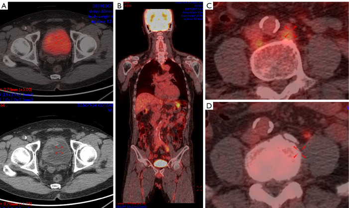
Figure 1.
A 65-year-old male with bladder cancer (A). Intense FDG-containing urine obscures FDG uptake in the bladder wall. Histopathologic analysis showed metastatic disease in a left paraaortic lymph node (B,C), which showed F-18 FDG uptake on a PET/CT, coronal, and axial PET/CT fusion image. After transurethral resection and chemotherapy, F-18 FDG avid lymph node enlargement disappeared (D).

