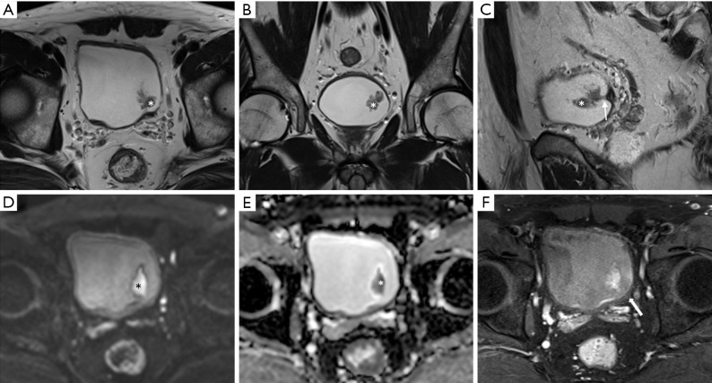
Figure 11.
Urothelial carcinoma (stage Ta) in a 56-year-old man. Axial (A), coronal (B), and sagittal (C) T2-weighted images showing an approximately 3-cm papillary mass (asterisk) in the left posterior wall. In axial and coronal images, the tumor appears to float on the bladder. However, the tumor stalk (arrow) is clearly observed in the sagittal image. A high b value diffusion-weighted image (D) and apparent diffusion coefficient map (E) showing the papillary mass with marked diffusion restriction (asterisk). There is no disruption of the muscle layer in the T2WI. Axial three-dimensional T1-weighted spoiled gradient echo image (F) obtained 180 seconds after contrast agent administration showing preserved mild delayed enhancement of the muscle layer (arrow). Therefore, the case can be classified as vesicle imaging-reporting and data system grade 2.

