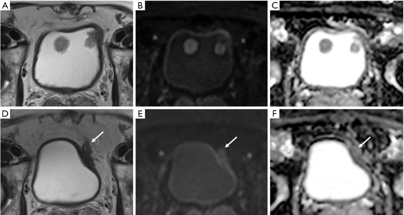
Figure 13.
Follow-up magnetic resonance imaging (MRI) of an 82-year-old man who underwent transurethral resection of bladder tumor (TURBT) for urothelial carcinoma. Axial T2-weighted image (T2WI) (A), high b value diffusion-weighted image (DWI) (B), and apparent diffusion coefficient (ADC) map (C) showing two masses in the bladder anterior wall. MRI shows a T2 hypointense bladder cancer without muscle invasion that was diagnosed as high-grade T1 urothelial carcinoma on TURBT specimens including proper muscle tissue. In follow-up MRI, axial T2WI (D), high b value DWI (E), and ADC map (F) obtained 8 months after TURBT showing new perivesical infiltration with hair-like projection (arrow) in the left anterior perivesical area. There was no remarkable diffusion restriction on DWI, and histopathological examination of repeat TURBT specimens containing proper muscle tissue showed acute and chronic inflammation in the adjacent bladder wall.

