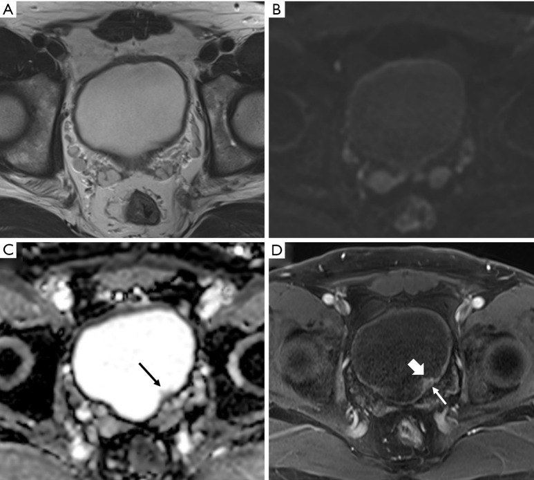
Figure 2.
Urothelial carcinoma (stage Ta) in a 74-year-old man. Axial T2-weighted magnetic resonance image (T2WI) (A) showing the normal bladder wall as a hypointense line. Axial diffusion-weighted image (DWI) with a high b value (B) showing the normal bladder wall as an intermediate signal intensity line. Axial apparent diffusion coefficient map (C) showing the bladder wall with low signal intensity. Small papillary lesion with intermediate signal intensity (arrow) in the left posterior wall that was not clearly visible on previous sequences. Axial three-dimensional T1-weighted spoiled gradient echo image (D) obtained 60 seconds after the administration of contrast material shows intense enhancement of the bladder tumor (thick arrow) unlike the muscle layer (thin arrow).

