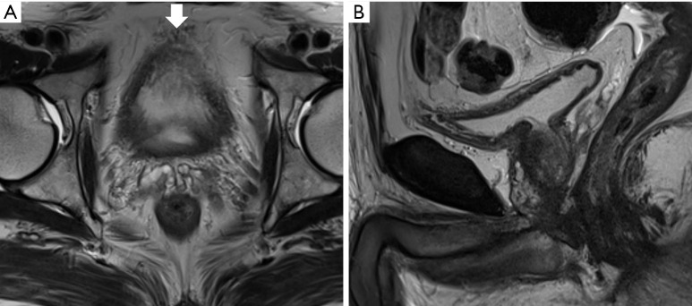
Figure 3.
Magnetic resonance imaging (MRI) of a 74-year-old man who underwent transurethral resection of a bladder tumor previously. Axial (A) and sagittal high-resolution T2-weighted magnetic resonance image (B) showing a poorly distended urinary bladder. The bladder lumen is barely visible on the axial image because of insufficient distension. The vascular structures (thick arrow) between the anterior and perivesical fat mimic tumor infiltration. Evaluation of the bladder wall and lumen on the sagittal image was limited yet marginally feasible; MRI recall or additional cystoscopy should be considered in such cases.

