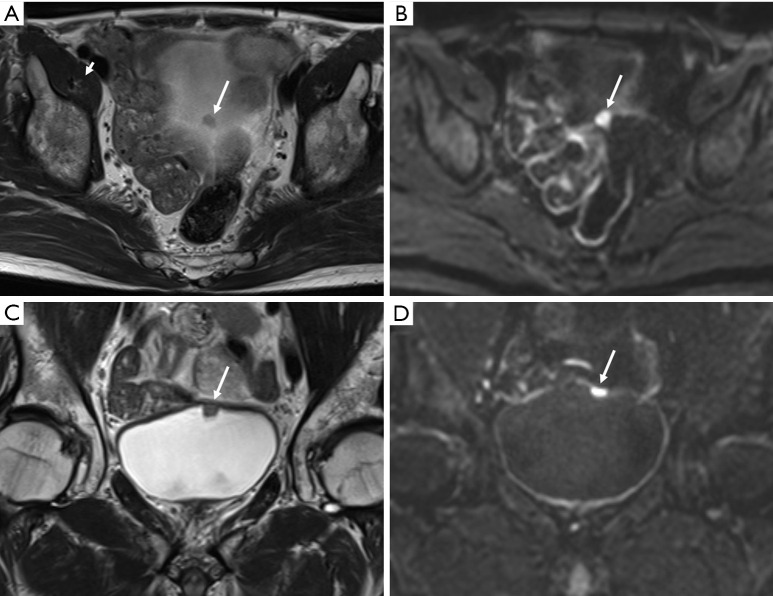
Figure 4.
Urothelial carcinoma (stage T1) in a 63-year-old man. Axial T2-weighted image (T2WI) (A) showing a small tumor in the bladder dome (arrow). In the diffusion-weighted image (DWI) with a high b value (B), the tumor shows markedly increased signal intensity (arrow). However, the localization of bladder tumors solely based on these images is difficult. The lesions located on the bladder dome are often not clearly visualized on axial images because of the persistence of partial volume artifacts. In such cases, coronal images (C, coronal T2WI; D, coronal DWI with high b value) are extremely helpful for tumor localization and T stage evaluation.

