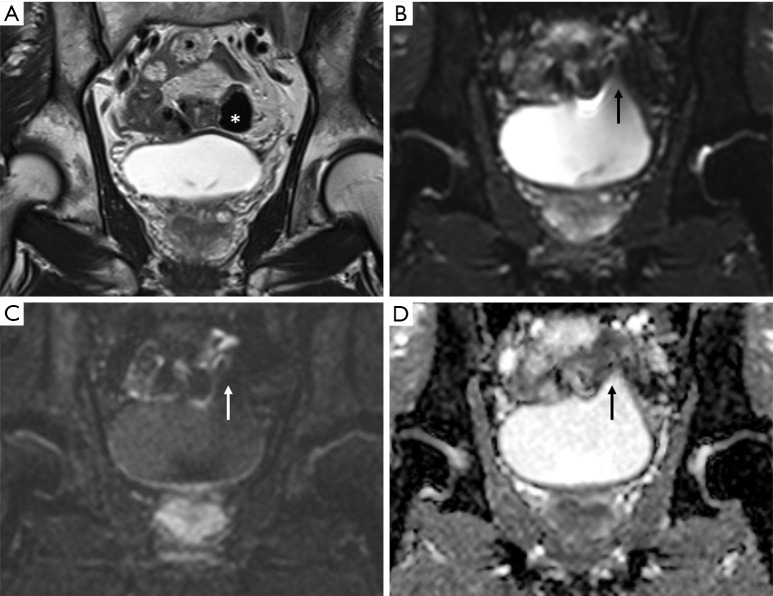
Figure 5.
Vulnerability on a coronal diffusion-weighted imaging. Additional slice of the coronal plane image of the patient described in Figure 4. (A) T2-weighted image (T2WI), (B) b =0, (C) b =1,000, and (D) apparent diffusion coefficient map. Coronal T2WI showing no abnormality in the left dome portion, and an air-filled bowel structure (asterisk) with dark signal intensity is located immediately adjacent to it. However, in the diffusion-weighted images, distortion is observed in the left dome of the bladder (arrow). It is believed that motion artifacts caused by bowel peristalsis and susceptibility artifacts caused by air existed simultaneously.

