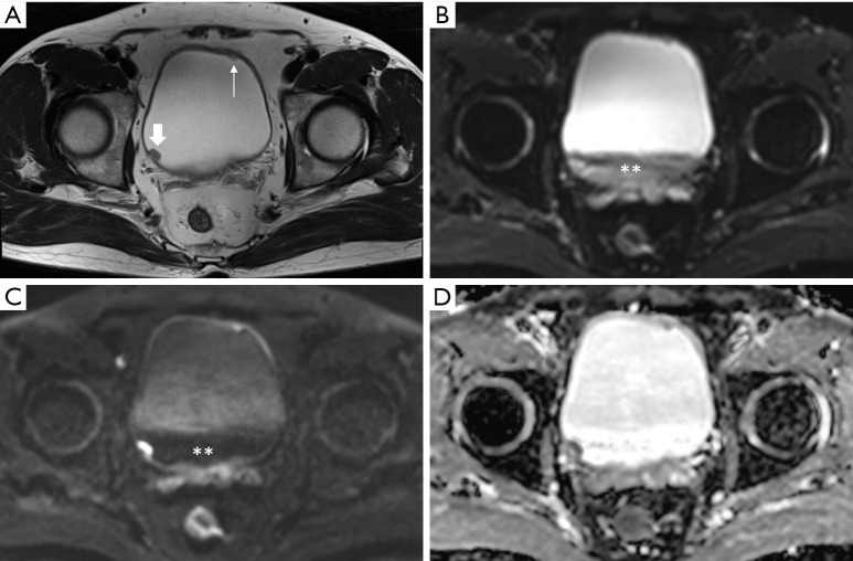
Figure 6.
Urothelial carcinoma (stage T1) in a 65-year-old man highlighting the importance of magnetic resonance (MR) image acquisition order. (A) A small tumor (about 1 cm) is visible in the right posterior wall (thick arrow), while another tiny polypoid lesion is seen in the left anterior wall (thin arrow) on an axial T2-weighted image. Diffusion-weighted image (B, b =0; C, b =800) and apparent diffusion coefficient map (D) obtained after delayed contrast enhancement shows a new heterogeneous low signal in the bladder-dependent portion (asterisk) that differs from the signal of urine in the nondependent portion. This represents the change in the signal intensity of urine because of excretion of the MR contrast agent that, to some extent, influences the contrast between the right posterior tumor and urine in the low b value image (B).

