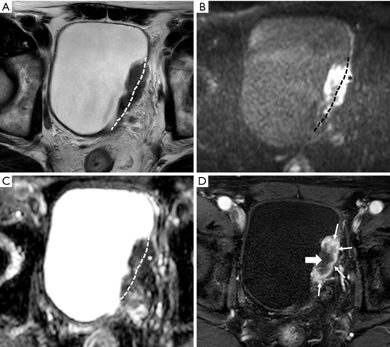
Figure 8.
Urothelial carcinoma (stage T3b) in a 79-year-old man. Axial T2-weighted image (A) shows a T2 heterogeneous mass with intermediate to low signal intensity in the left wall. If the dotted line is considered the boundary between the bladder wall and the perivesical fat, a tumorous lesion extending into the perivesical fat area is observed, which can be staged as T3b. A high b value image (B) and apparent diffusion coefficient maps (C) also show diffusion restriction in the tumors extending to the bladder wall (asterisk). Axial three-dimensional T1-weighted spoiled gradient echo image (D) obtained 40 seconds after contrast agent administration shows early peripheral enhancement (thin arrow) and central necrosis (thick arrow) of the large tumor. This case is classified as vesicle imaging-reporting and data system grade 5.

